Top 5 Places to Perform Skin Scrapes on a Koi Fish
Posted by Ellen Kloubec on 8th Sep 2014
The purpose of taking a skin scrape is to obtain a sample of mucus from koi’s cuticle, or slime coat, to analyze under a microscope. The mucus specimen is examined for significant parasitic existence and identification, and as a precursor to treatment. Parasites are always present in low numbers but when koi become stressed parasites will flourish, become problematic, and can lead to bacterial infections. Performing a skin biopsy, or skin scrape with a glass slide or coverslip can be intimidating, but is the best method to accurately diagnose parasite existence and type. Getting good quality skin scrapes will take practice and patience.
WHERE TO SCRAPE:
- 1.Flank: Along the fish’s side above lateral line.
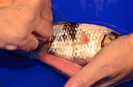
The easiest place to obtain a respectable mucus sample is from the flank of the koi. Begin at the shoulder and drag a glass slide towards the tail. You should accumulate ample mucus for your sample.
- 2.Caudal: Body onto tail.
The caudal region is the second easiest area for performing skin scrapes. Starting midway on the body, below the lateral line, scrape towards the tail, ending at mid-tail.
- 3.Gill Operculum: External gill cover and onto the pectoral fin.
Scrapes taken from the gill cover and onto a pectoral fin will be very productive when searching for parasites, especially flukes. Place the edge of a glass slide on the gill cover (operculum) and scrape downwards and onto the pectoral fin.
- 4.Chin: Cleft between gill covers underneath the fish.
You will need to roll the koi on its back to obtain a scrape from underneath the chin or cleft between the gill covers. This will be a ‘short-but-sweet’ scrape that is usually abundant with parasitic organisms. Scrape the cleft and onto the pectoral fin joint.
- 5.Wound or Ulcer: A sore or any spot that shows damage.
If your koi has a lesion or ulcer you can bet that a biopsy of that location is sure to be rich with parasites. Scrape the entire area around the wound for an accurate picture of the extent of parasite infestation.
Always scrape in the direction from head to tail, or with the scales. Do not go against the scales as you may damage the koi. Keep prepared slides out of direct sunlight. For best results your samples should be observed under a microscope within 30-60 minutes after being collected.


